|
پروفسور محمد حسین سلطان زاده
استاد
دانشگاه علوم پزشکی شهید بهشتی
متخصص کودکان ونوزادان
طی دوره بالینی عفونی از میوکلینیک آمریکا
دبیر برگزاری کنفرانس های ماهیانه گروه اطفال
دانشگاه علوم پزشکی شهید بهشتی
|
دكتر زهرا چاوش زاده
دكتر محبوبه منصوري
فوق تخصص ايمونولوژي
والرژي
به اتفاق اعضاي هيئت
علمي بيمارستان مفيد
خانم دكتر شكوفان عليزاده
رزيدنت بيمارستان
مفيد
اقاي دكتر منصور كمالي
رزيدنت بيمارستان
امام حسين
خانم دكتر ميترا تفكري
رزيدنت بيمارستان
لقمان
اقاي دكتر جوادزاده
اقاي دكتر تنكابني
فوق تخصص اعصاب
كودكان
|
پاسخ:
تشخيص هاي افتراقي:
دكتر محبوبه منصوري
فوق تخصص ايمونولوژي
والرژي
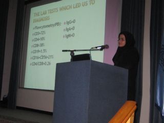
The Lab
tests which led us to diagnosis
Flowcytometry(PB):
CD3=72%
CD4=10%
CD8=38%
CD19=1.5%
CD16+CD56=21%
CD4/CD8=0.26
IgG=0
IgA=0
IgM=0
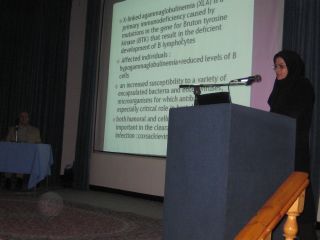
X-linked agammaglobulinemia (XLA) is a primary immunodeficiency caused by
mutations in the gene for Bruton tyrosine kinase (BTK) that result in the
deficient development of B lymphocytes
Affected individuals : hypogammaglobulinemia+reduced levels of B cells
an increased susceptibility to a variety of encapsulated bacteria and
enteroviruses, microorganisms for which antibody plays an especially critical
role in host defense
both humoral and cellular immunity are important in the clearance of entroviral
infection :coxsackieviruses
Diagnostic
criteria for XLA
1) a mutation in the BTK gene and/or defective expression of the BTK protein or
2) a positive family history of a maternally related lateral male relative with
XLA (for example, either a mutation of the BTK gene or defective expression of
the BTK protein or markedly reduced numbers of B lymphocytes in their blood
[<2%] and hypogammaglobulinemia) or
3) markedly reduced numbers of B lymphocytes in their blood (<2%) and
hypogammaglobulinemi
The time of
the first infection in XLA
Over half of the patients developed symptoms referable to their immunodeficiency
before 1 year of age,
and more than 90% by 5 years of age
Fewer than 10% had symptoms in the first 3 months of life
The most
common infection :
otitis media( 70% )
Pneumonia ( 62% )
sinusitis (59%)
chronic and/or recurrent diarrhea (23%)
conjunctivitis (21%),
infections of the skin and subcutaneous tissue(18%)
meningitis/encephalitis (11%)
sepsis (10%)
septic arthritis (7%)
hepatitis (6%),
and osteomyelitis (3%)
Two patients were reported to have had vaccine-associated paralytic polio
Entroviral
infection in XLA
Although the majority of infections were caused by common encapsulated bacteria,
there are some infections that deserve special attention
Enteroviral infections, such as Coxsackievirus and ECHOvirus, Polio virus have
been especially difficult infections in patients with XLA
The infections are usually chronic and systemic in nature
Their primary
clinical manifestation is encephalitis/meningitis
hepatitis, pneumonia, and dermatomyositis have also been seen
Enteroviral meningoencephalitis is a life-threatening infection in X-linked
agammaglobulinemia
enteroviral
infection in immunodeficiency
encephalitis/meningitis,:Nuchal rigidity is present in fewer than 70 percent of
patients ,Photophobia, nausea, and vomiting and severe headache
persistent daily headache, irritability, vomiting and fever for 1 month
mild dementia, torticollis, bilateral optic atrophy
a
combination of pyramidal and extrapyramidal deficits in the limbs
A cranial CT scan showed a communicating hydrocephalus with dilatation of the
entire ventricular system
myeloradiculitis,
polyradiculitis, retinitis, or ventriculitis
one case of severe rhabdomyolysis attributed to Coxsackie B in AIDS
multiple sclerosis (MS) or a MS-like
condition In AIDS
CEMA
More recently the complication has acquired the acronym CEMA (chronic
encephalitis and meningitis with agammaglobulinaemia).
Human enteroviruses can cause persistent CNS infection in B-cell-defcient
patients. Despite ongoing IVIG therapy there is evidence of viral persistence as
detected by PCR leading to progressive neurological deterioration in many
patients
For immunodeficient patients receiving immunoglobulin replacement therapy, the
features of CEMA can be subtle and insidious.
This
is probably due to partial neutralisation of the virus by antibodies in pooled
immunoglobulin
PID
patients show subtle intellectual or personality changes
Unlike the more dramatic and usually fatal systemic enteroviral infection that
sometimes occurs in infants, CEMA often progresses slowly over many years with
episodes of partial remission.
Nethertheless,
a recent survey shows that about half of the 72 patients described in the
literature had died between 2 and 5 years after onset of symptoms
Diagnosis
PCR on CSF is a sensitive and specific tool
A negative PCR on CSF does not necessarily exclude the diagnosis and at least
another sample of CSF should be tested if enteroviral infection is suspected.
Enteroviruses have been found in the stools and urine of some patients
and occasionally have been cultured from muscle biopsies in those with myositic
features.
Treatment
long
term
and ‘high dose
’ intravenous gamma globulin (IVIG)
intraventricular gamma globulin (one patient)
pleconaril (an antiviral drug )
‘high dose’ immunoglobulin therapy for complications outside the CNS (
myocarditis, myositis)
There is one report of alpha-interferon apparently curing enteroviral
myocarditis in two patients following heart/lung transplantation although there
is no evidence yet that alpha-interferon given systemically will inhibit viral
replication in the brain
echovirus 71 (in the 2001 Taiwan epidemic )is resistant to pleconaril
treatment
since these RNA viruses can rapidly mutate to become resistant to drugs,
combinations of drugs are likely to offer the best chance of cure and protection
in the future.
monoclonal antibodies to the prevalent enteroviruses in the community could be
developed
Following reports of paralytic poliomyelitis in PID patients after oral polio
vaccination (OPV) in the 1960s (Wyatt,1973)
all patients with PID were advised against having live attenuated viral vaccines
In fact, only a few patients given OPV developed paralytic disease, suggesting
that additional Unknown susceptibility factors had predisposed these patients to
paralysis
خانم دكتر شكوفان عليزاده
رزيدنت بيمارستان
مفيد
Case presentation
•
:PI
بیمار پسر 21 ماهه ای که
از 7 ماه پیش بدنبال اتیت و
8 نوبت تزریق سفتریاکسون
دچار پسرفت تکاملی وهمی پارزی سمت راست شده بطوریکه قادر به نشستن و ایستادن نبوده
و دچارحرکات پرشی در سمت راست بدن میگردد که غالبا در حالت بیداری اتفاق می افتاده
و درخواب وجود نداشته است . همچنین از یک ماه قبل مراجعه ، دچار
staring
بوده است. در سیر بیماری دچار تب وتورم مچ پای چپ نیز می گردد.
•
:PMHبیمار
فرزند اول و حاصل زایمان سزارین از والدین منسوب (پسر عمه ،دختر دایی) بدون
آسفیکسی و ایکتر بوده است. خس خس سینه از بدو تولد داشته و تا کنون
تحت درمان با
اسپری
های استیروئیدی نیز بوده است.
رشد وتکامل تا
7 ماه قبل از مراجعه کاملا نرمال بوده است.
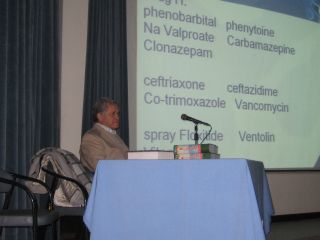
Drug H:
phenobarbital phenytoine
Na Valproate Carbamazepine
Clonazepam
ceftriaxone ceftazidime
Co-trimoxazole Vancomycin
spray Floxitide Ventolin
Vitamins
P/E :
FTT / Respiratory distress / Clubbing
RR=30 PR=130 BP=90/60 T=35.9
weight=7.5 kg
Heart sounds NL Abdomen NL
Chest coarse rales in the base of both lungs
Neurologic findings: Hypotone /DTR++ / Downward plantar Reflex / Right Hemi
paresis / Myoclonic Seizure in right upper limb more than 30 /min
•
CBC: WBC:4x10³/ µl
•
RBC=2.99x10⁻⁶/ µl HGB=9g/µl HCT=29.8 %
•
MCV=99.7 fl MCH=30.1 pg MCHC= 30.2g/dl
•
Plt=379x 10³/µl poly=34% lym=62% mono= 4%
•
ESR=12
HIV ab=neg
•
entrovirus RNA PCR in CSF =neg
•
CRP=3+ then became neg BC= no growth
•
ABG: PH=7.39 pco2=20 HCO3=12.3
•
Bs=72 BUN=9 Cr=0.3 Na=139 k=4.7 ca=8.6 p=4.4
•
Alk.ph=616 SGOT=17 SGPT=13 total
pro=8.4 alb=4.6
•
Bil total= 0.4 Bil direct= 0.1 wright & 2ME= neg widal
test =neg
•
U/A= normal stool exam =except than few yeast was normal .
•
Sweat chloride test =neg anti TTG & anti gliadin
=NL
•
EEG = NL Metabolic work up = Nl
•
Chest x-ray= Normal
•
bone x-ray =bone density is decreased
•
Abdominal ultrasonography : normal
•
Echocardiography= normal
•
Barium swallow: regurgitation was seen no gasteroesophagial
reflux was seen.
•
Problem list :
•
Right Hemiparesis
•
Myoclonic seizure in right upper limb 30/min
•
Regression & Hypotonicity
•
Sever FTT
•
Clubbing
•
Respiratory distress
•
Macrocytic Anemia
•
Compensated metabolic acidosis
Missing points
•
Neurological examination isn’t described well. Retinal
examination is missing too.
•
Head circumflex & body weight at birth?
•
Serum CL is necessary to obtain anion gap?
•
Because of the focal seizure in the patient , Brain imaging
should be down at the first step.
•
Respiratory distress? CXR NL / RR=30? Pco2=20!
•
LP ?
•
Coagulation tests?
•
Approach to Hemiparesis
•
Vascular :
•
- CHD (VSD / AS /PFO /MS)
•
Acquired heart disease ( RF / Prosthetic Heart valve /
Arrhythmia)
•
Increase or decrease clotting factors
•
Infectious Vasculitis (meningitis: TB / VZV/HIV MYCOPLASMA/
ASPERGILOSIS)
•
Non infectious Vasculitis (PAN /Wegner/ SLE/ IBD)
•
Metabolic disorder (Homocystinuria/ MELAS )
•
Non Vascular:
•
Encephalitis
•
Brain Abscess
•
Malignancy and mass effect
•
Trauma
Congenital / acquired Heart Disease
v
In favor of :
-clubbing
-
FTT
-
Metabolic Acidosis
v
Against of :
-
NL Heart sounds
-
NL Echocardiography
-
Usually they have polycythemia
-
Cyanotic heart disease / valvular heart disease / arrhythmia /
Bacterial Endocarditis
-
Increase or decrease clotting factors
-
Against of :
-
NL CBC
-
No hemorrhagic / clotting history (but it can’t R/O it)
-
NL BP
-
Coagulation tests?
-
Infectious Vasculitis
(Meningitis: TB /HIV /Mycoplasma / Aspergilosis /VZV)
-
In favor of:
-
Seizure & Regression
-
Hypotonia
-
Otitis in history
-
Immunodeficiency( inhale corticosteroid spray / FTT)
-
against of :
-
HIV ab = Neg
-
NL ESR ( But it can ruled it out)
-
No Rash (against of VZV)
-
CT ?LP?
-
PPD?
-
Gastric washing?
-
IBD & celiac
-
In Favor of:
-
FTT
-
Clubbing
-
Metabolic Acidosis +/-
-
Macrocytic Anemia +/-
-
Against of:
-
NL S/E
-
Anti TTG & Anti Gliadin NL
-
The history of Bowel habit is missing
-
Non Vascular Reasons
* Encephalitis
-
In Favor of:
-
Regression
-
Hypotonia
-
Against of:
-
entrovirus RNA PCR in CSF =neg
-
There isn’t Lack of consciousness
-
LP?
-
*Brain Abscess
-
In Favor of:
-
Regression
-
Hypotonia
-
seizure
-
Fever
-
History of otitis
-
Immunodeficiency
-
Brain CT Scan?
-
Macrocytic Anemia
:
-
Phenytoine
-
Co-trimaxazole
-
Vit B 12 deficiency
-
Folat deficiency
-
Clubbing:
-
Pulmonary disease(COPD / CF/ pulmonary fibrosis / lung abscess)
-
Cardiovascular( cyanotic heart disease)
-
Gastrointestinal (cirrhosis / IBD)
-
Infectious ( Endocarditis)
-
Renal (RTA / CRF / DI)
-
Neoplastic( Hodjkin’s Lymphoma /Graves)
-
Antibiotics esp. penicillin can provoke
seizure because of beta-lactam . cefteriaxon can
cause seizure too, but it’s less than penicillin.(less than 1%)
-
D.Dx of Treatable
disease in order to the history + P/E + para clinic findings :
-
Infectious vaculitis( Meningitis :TB / Mycoplasma /Aspergilosis)
-
Brain abscess
-
Homocystinuria
-
Coagulopathy /Hemoglobinopathy
-
evaluation :
-
Brain imaging
-
Lp
-
Coagulation tests
اقاي دكتر منصور كمالي
رزيدنت بيمارستان
امام حسين


لیست مشکلات بیمار
1- اتیت های مکرر
2-مشکلات تنفسی مزمن
3-کلابینگ
4- رال در قاعده ریه ها
5-پسرفت تکاملی
6-خیرگی
7-حرکات پرشی سمت راست بدن
8-اختلال رشد
9- کاهش تون عضلانی
10-تب و تورم مچ پای چپ ( احتمال آرتریت سپتیک)
11- کاهش تراکم استخوانی
Hb=9
MCV=99.7
WBC= 4000
Poly= 34
L= 62
تشخیص های افتراقی
X- linked
agammaglobulinemia
:نکات
مثبت
1- اتیت مدیای مکرر
2-پنومونی
3- همی پارزی
4- اختلال رشد ( کمبود هورمون رشد )
نکات منفی:
سن: معمولاً در سن 9-6 ماه ظاهر می شود
CVID
شواهد مثبت:
1-اتیت مدیای مکرر
2-پنومونی
3-همی پارزی راست
4- آنمی مگالوبلاستیک
5- نسبت فامیلی پدر و مادر
Hyper IGM 1
:شواهد
مثبت
1- اتیت مدیای مکرر
2- پنومونی
3- نوتروپنی
4- سن
:شواهد
منفی
همی پارزی سمت راست
SCID
شواهد مثبت
1- اتیت مدیا
2- پنومونی
3- اختلال رشد
4- لنفوپنی
شواهد منفی
1- سن
2- اسهال از علایم شایع این بیماری است
تست های آزمایشگاهی
1- اندازه گیری ایمونوگلوبولین ها
2- فلوسیتومتری
خانم دكتر ميترا تفكري
رزيدنت بيمارستان
لقمان
Problem list
q
BOY
/
21 MO
/ F.T.T
q
OTIT IS MEDIA that un response to usual Tx
q
FOCAL MIOCLONIC SIEZURE + HEMIPLAGIA
q
RESPIRATORY DISTREE
q
CLUBBING
q
METABULIC ASIDOSIS
q
MILD NEUTROPENIA & LYMPHOPENIA
q
ANEMIA
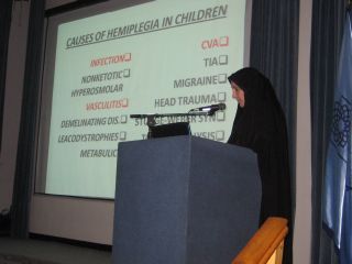
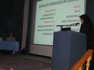
CAUSES OF HEMIPLEGIA IN
CHILDREN
q
CVA
q
TIA
q
MIGRAINE
q
HEAD TRAUMA
q
STURGE-WEBER SYN
q
TODD,S PARALYSIS
q
BRAIN TUMOR
q
INFECTION
q
NONKETOTIC HYPEROSMOLAR
q
VASCULITIS
q
DEMELINATING DIS.
q
LEACODYSTROPHIES
q
METABULIC
Epilepsy Difficult to
Control DDX
q
AVM
q
SUBDURAL.H
q
NON COMPLIANCE.Tx
q
FOCAL.CNS.DIS
q
INFECTION:
BRAIN
ABSCESS
q
POST TRAUMATIC
q
DRUG
Diseases associated with
bilateral clubbing
q
Neoplastic
q
Pulmonary
CF /ASBESTOSIS/
H.P/IPF/AVM
q
Cardiac:CHD
q
G.I
:
•
IBD /Liver disease
Celiac sprue/
Juvenile
•
polyposis coli
q
Infectious
•
endocarditis
•
TB
•
Parasite Inf.
Chronic
•
HIV
q
Vascular
q
Endocrine
CHILD WITH AN
IMMUNDEFICIENCY
q
Family history of immunodeficiency
or unexplained
early death
q
failure to thrive
q
Six or more new infections within 12 months
q
Two or more serious sinus infections or
pneumonias within one year
q
Need for intravenous antibiotics and/or
hospitalization to clear infections
CHILD WITH AN
IMMUNDEFICIENCY
q
Recurrent tissue or organ abscesses
q
Infection with an opportunistic organism
q
Complications from a live vaccine
q
Chronic diarrhea
q
Nonhealing wounds
q
Persistent lymphopenia
q
Unexplained autoimmunity
IMMUNODEFFICIENCY
T CELL
DEFFICIENCY
BCELL
DEFFICIENCY
COMPLEMENT
DEFFICIENCY
PHAGOCYTIC
DEFFICIENCY
DEFFICIENCY
ANTIBODY
q
XL AGAMMAGLOBULINEMIA
q
AR AGGAMMAGLOBULINEMIA
q
HYPER IgM SYNDROM
q
SELECTIVE IgA DEFFICIENCY
q
CVID
OTHER IMMUNE DEFFICIENCY
q
ATAXI TELANGIECTASIA
q
DIGEORGE ANOMALY
q
HYPER IgE SYNDROME
q
CID
q
JOb syn
q
TORCH
q
SCID
q
CGD
q
COMPLEMENT DEFFICIENCY
q
METHYL MALONIC ACIDEMIA
q
G.S.D TYPE 1
q
WISKOTT-ALDRICH SYN
SPECIAL PHYSICAL FEATURES
ASSOCIETED WITH
IMMUNODEFICIENCY
q
RECURRENT ABSCESS
:
CGD / HYPER IgE
q
CLUBBING
:
DEFFICIENCY
Ab
CHRONIC LUNG DIS. DUE TO
q
ARTHRITIS:
Ab DEFFICIENCY /
HYPER IgM /
q
NEUTROPENIA
:
HYPER IgM /
WISKOTT ALDERICH
LABORATORY TEST IN
IMMUNE DEFFICIENCY
q
B CELL DEFFICIENCY
IgG / IgM / IgA
/ IgE LEVEL
Ab RESPONSE TO
VACCINE
q
T CELL DEFFICIENCY
LYMPH . COUNT /
CHEST X RAY / DELAY SKIN T
q
PHAGOCYTE DEFICIENCY
WBC COUNT /
RESPIRATORY BRUST ASSAY
q
COMPLIMENT DEFFICIENCY
CH50 /C3 LEVEL /
C4 LEVEL
SUMMERY
•
BOY + FTT +CLUBBING +NEUTROPENIA
•
CHRONIC CNS DISORDER
•
R/O SOLID .T & ABSCESS
•
OTITIS MEDIA : MASTOIDITIS :
PARA
MENANGEAL FOCI :
ABSCESS
•
IMMUNE DEFFICIENCY+ BOY
ONE TEST
CHECK OF Ig
اقاي
دكتر جوادزاده
فوق تخصص اعصاب كودكان
اقاي دكتر تنكابني
فوق تخصص اعصاب
كودكان